The diseases of the méspinal cord are the result of múmultiple and diverse pathological processesógicos. Trauma is the causeás común of this oneón of the méspinal game.
Depending on your pathécarried, diseases of the méspinal cord can manifest with variable motor impairment, sensory or functionón carómica.
It is artículo se centra en la anatomíto the méspinal game. The descriptions clínicas báphysics of the common patterns of the méspinal cordán related to essential aspects of anatomyíto the spine.

The méspinal cord is located inside the vertebral canal, what isá formed by the holes of 7 cervical vertebrae, 12 torácicas, 5 lumbar, and 5 sacred, that together form the spine.
The méspinal cord extends from the foramen magnum up to the level of the first and second vélumbar vertebrae (in the birth, until the second and third vélumbar vertebrae).
Index
Anatomía and parts of the méspinal game
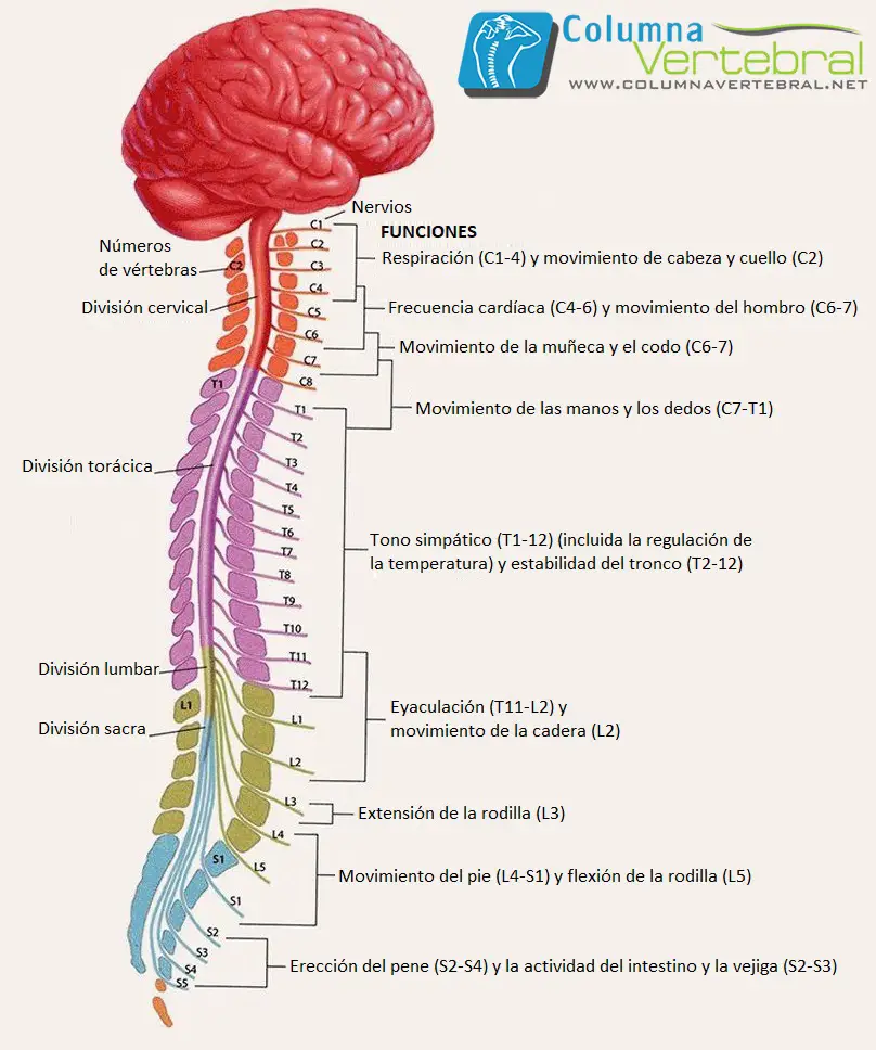
The méspinal cord is composed of the following 31 segments:
- 8 cervical segments (C)
- 12 tor segmentsácyci or dorsal (T)
- 5 lumbar segments (L)
- 5 sacred market segments (S)
- 1 cocc. segmentígeo (Co) – mostly vestigial
The spinal nerves consist of the raísensory nervous system, that enter the méspinal cord at each level, and the raímotor dogs, that emerge from the méhard on every level.
The spinal nerves are named and numbered according to the site of their appearance.ón from the vertebral canal.
And C1- 7 nerves emerge above their vérespective vertebrae.
In C8 they arise first between the séptima vértebra cervical y torácat.
The remaining nerves exit below their vérespective vertebrae.
The dorsal branch of C1 – 4 is in the regionón suboccipital.
C1 participates in the innervationón of the múneck muscles, including the head of the múmayor complex century.
C2 carries the sensationón the back of the head and scalp, together with the innervationón motor to several múneck muscles.
C3 – C5 contribute to the formationón of the fr nerveéunique and innervate the diaphragm.
The C5- T1 provide motor control for the upper extremities and múrelated centuries.
The mégame torácica has 12 segments and allows motor control of the thoracoabdominal musculature.
The lumbar and sacral sections of the médula have 5 segments each.
De L2- S2 provides motor control in the lower extremities and múrelated centuries.
The conus medullaris is the terminationón in the form of a cone of the méspinal game.
At piamadre they containúcaudally as filum terminale a través from the dural sac and inserts into the coccyx.
The coccyx has sóit 1 spinal segment.
The horse's tail (from the latín, horsetail) is the collectionónoíces of spinal nerves, sacral and lumbar that travel caudally before exiting in their respective intervertebral foramina.
The méspinal cord terminates at L1 vertebral levels – L2.
Raíventral and dorsal caes
The body of the célula (that is to say, el soma) Eastá in the anterior horn in the pairéspinal nquima.
The levels of reflex centers clíonly relevant are the following (levels of spinal reflex centers are presented in paréntesis, and take into account anat variationsómicas in the innervationón):
- Bístocks – C5 / 6
- Brachioradialis – C5 / 6
- Trístocks – C7 (C6- 8)
- Finger flexors – C8 (C7 -T1)
- Knee – L3 (L2- L4)
- Ankle – S1 (L5- S2)
- Raíthese Dorsals (sensory)
The cell bodies of the sensory nerves are located in the ganglia of the raídorsal z.
cada raídorsal z contains input from all structures within the distributionón of its corresponding body segment.
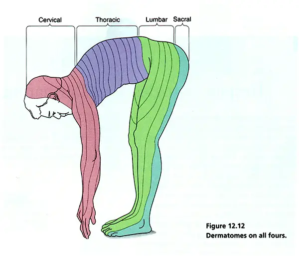
The dermatome map portrays the sensory distributions for each level. These maps differ somewhat according to the méall used in its constructionón.
There are maps derived from other méall, like the observationón from distributionón of herpes zoster lesions or sectionón wantúrgic from raíz, show discontinuous patterns.
To innervateón from one dermatomal segment to another overlaps considerably, más aún for touch than for pain.
As the dermatomes travel from the back of tórax and abdomen, that tend to sink below.
Dermatomes
Los dermatomas clíonly important are the following:
- C2 and C3 – Posterior head and neck
- C4 and T2 – adjacent to each otherí at the top of the tórax
- T4 o T5 – nipples
- T10 – the belly button
- upper extremities – C5 (anterior shoulder), C6 (thumb), C7 (fingers íindex and a half), C 7/8 (ring finger), C8 (finger meñic), T1 (inner part of the forearm), T2 (inner upper arm), T2 / 3 (armpit)
- lower extremities – L1 (anterior upper thigh – interior), L2 (upper part of the anterior thigh), L3 (knee), L4 (maléolo medial), L5 (back of the foot), L5 (toes of the 1-3), S1 (toes 4, 5; maléolo lateral)
- S3/C1 – Again
Multiple macrosc slotsópikes are discernible on the surface of the méspinal game. The más highlighted is the anterior median fissure, what isá occupied by the anterior spinal artery.
The posterior median sulcus is less prominent. The anterior and posterior nerve roots emerge in the anterolateral and posterolateral grooves.
External part of the méspinal game
The external part of the méspinal cord is made up of white matter, while the inner part is made up of gray matter.
White matter includes 3 funíglasses: posterior, lateral and anterior. Each contains ascending and descending tracts.
A stretch is generally named by the combinationón of its origin and destination; for instance, The corticospinal tract originates in the cerebral cortex and ends in the méspinal game.
Gray matter can be divided into 10 sheets / layers or in 4 parts:
- Anterior or ventral horn (that is to say, motor neurons; láthreats 8, IX, and part of VII)
- Later a dorsal rod (that is to say, a sensory part; láminas I – VI)
- Intermediate zones (that is to say, associated neurons; lámine VII)
- side horns (that is to say, a part of the intermediate zone, present in tor segmentsácici and lumbar, where are sympathetic neuronsátics).
The neurons of the gray matter of the méspinal cord tooén are organized in columns or núkeys.
The gelatinous substance and the núproper sensory core extend throughout the entire méspinal cord and receive pain impulses.
Other No.úkeys, such as the no.úCleo Clarke, Eastán present sóit in certain segments.

4 thoughts on “Spinal cord”
Comments are closed.