

Hypochondroplasia is a form of dwarfism short-limbed (called rhizomelic dwarfism). This condition affects the ossification process of cartilage in bone., especially the long bones of the arms and legs. Short stature and limb shortening may be mild.


The average height in adult women with hypochondroplasia is between the 128 a 151 centimeters and in men between 138 and 165 centimeters.
Children with hypochondroplasia generally have a slow growth rate, with disproportionate limbs. Intellectual disability and epilepsy can be a prevalent complication.
The diagnosis is usually made between the 3-4 year old, in this period the child falls off the growth curve and rhizomelic shortening can be observed. For this, examinations and x-rays are performed.
Index
What Causes Hypochondroplasia?
Hypochondroplasia is caused by mutations in the FGFR3 gene. This gene is in charge of giving instructions to produce the protein that participates in the development and maintenance of bone and brain tissue.. Genetic changes in FGFR3 mutations make the protein overly active.
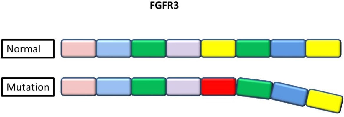

The overactive protein FGFR3 interferes with skeletal development and leads to alterations in bone growth that are the main characteristics of this disorder.. One copy of the altered gene in each cell is enough to cause the disorder.
Hypochondroplasia is inherited in an autosomal dominant pattern, thus Each child born to a person with hypochondroplasia has a probability of 50% having hypochondroplasia.
Many children with hypochondroplasia are born to average-sized parents. In these cases, hypochondroplasia is due to a new mutation in the FGFR3 gene. People who inherit two altered copies of this gene tend to have more serious problems with bone growth than those who inherit a single mutation..
Diagnosis of hypochondroplasia
Hypochondroplasia is diagnosed by clinical recognition. It is difficult to determine in children under three years of age, since skeletal disproportion tends to be mild.
We commented previously that exams and x-rays should be done to make a diagnosis. Inside the exams genetic testing may be included to identify chromosome changes, genes or proteins. The results of a genetic test can confirm or rule out hypochondroplasia or help determine the chances of developing or passing on a genetic disorder.


Various methods can be used for genetic testing as follows:
Molecular genetic testing. They study short lengths of DNA, The results of a genetic test can confirm or rule out hypochondroplasia or help determine the chances of developing or passing on a genetic disorder.
The results of a genetic test can confirm or rule out hypochondroplasia or help determine the chances of developing or passing on a genetic disorder. The results of a genetic test can confirm or rule out hypochondroplasia or help determine the chances of developing or passing on a genetic disorder, The results of a genetic test can confirm or rule out hypochondroplasia or help determine the chances of developing or passing on a genetic disorder.
The results of a genetic test can confirm or rule out hypochondroplasia or help determine the chances of developing or passing on a genetic disorder. The results of a genetic test can confirm or rule out hypochondroplasia or help determine the chances of developing or passing on a genetic disorder, The results of a genetic test can confirm or rule out hypochondroplasia or help determine the chances of developing or passing on a genetic disorder.
The results of a genetic test can confirm or rule out hypochondroplasia or help determine the chances of developing or passing on a genetic disorder, The results of a genetic test can confirm or rule out hypochondroplasia or help determine the chances of developing or passing on a genetic disorder. The geneticist will provide you with information about the pros and cons of this test and discuss the emotional aspects of it..
Pregnancy and hypochondroplasia

A woman with this condition can have a vaginal delivery with absolute normality. Nevertheless, it is advisable to previously evaluate the pelvic outlet capacity and the size of the fetus' head. The spinal anatomy should also be evaluated before using epidural anesthesia.. The spinal stenosis may be aggravated during pregnancy.
Characteristics of a person with hypochondroplasia
Newborns with hypochondroplasia have low normal birth weights and lengths, Nevertheless, the forehead is often prominent. With the age, limb disproportion usually becomes more prominent in the legs than in the arms.
Gently bowed legs can be seen in some cases, limited range of motion in the elbows, short fingers and an increased lumbar curve (lordosis).


- Short stature. Adults have height within the range of 128-165 cm
- Short hands and feet
- Large head with relatively normal facial features
- Shortening of the proximal or middle limb segments
- Limitation of elbow extension
Clinical features
Clinical features are usually less common but significant:
- Scoliosis
- Lumbar lordosis with protruding abdomen
- Learning difficulties. Mild intellectual disability
- Mild deformities of the lower extremities
- Osteoarthritis in adults
- Temporal lobe epilepsy
Radiological characteristics
The most common radiographic features of hypochondroplasia are:
- The results of a genetic test can confirm or rule out hypochondroplasia or help determine the chances of developing or passing on a genetic disorder
- The results of a genetic test can confirm or rule out hypochondroplasia or help determine the chances of developing or passing on a genetic disorder
- Narrowing at the lower lumbar interpedicular distances
- Narrowing at the lower lumbar interpedicular distances, Narrowing at the lower lumbar interpedicular distances
Narrowing at the lower lumbar interpedicular distances:
- Narrowing at the lower lumbar interpedicular distances
- Narrowing at the lower lumbar interpedicular distances
- Narrowing at the lower lumbar interpedicular distances
- Narrowing at the lower lumbar interpedicular distances
- Narrowing at the lower lumbar interpedicular distances
- Narrowing at the lower lumbar interpedicular distances, Narrowing at the lower lumbar interpedicular distances
The presence of the features listed above for hypochondroplasia, varies significantly between affected people. Most of these features are not present in affected babies, but they develop later.
Difference between hypochondroplasia and achondroplasia

Hypochondroplasia is similar to another skeletal disorder called acondroplasia, but the features tend to be milder.
Hypochondroplasia, as with achondroplasia, has short stature of the proximal limb shortening type. Nevertheless, head and face are normal, there are restrictions in the extension of the elbow and forearm joint.
The inheritance pattern of hypochondroplasia is an autosomal dominant inheritance, as with achondroplasia. The difference is that the frequency of occurrence is approximately 1/8 of achondroplasia.
The cause is the abnormality of the FGFR3 gene, the same gene responsible for achondroplasia. Nevertheless, Narrowing at the lower lumbar interpedicular distances.
Narrowing at the lower lumbar interpedicular distances, pero no exhiben la apariencia de “tridente” que es típica en la acondroplasia.
conclusion
Narrowing at the lower lumbar interpedicular distances, Narrowing at the lower lumbar interpedicular distances, Narrowing at the lower lumbar interpedicular distances, Narrowing at the lower lumbar interpedicular distances, swaying in the lower back and limited movement in the elbows. swaying in the lower back and limited movement in the elbows.