


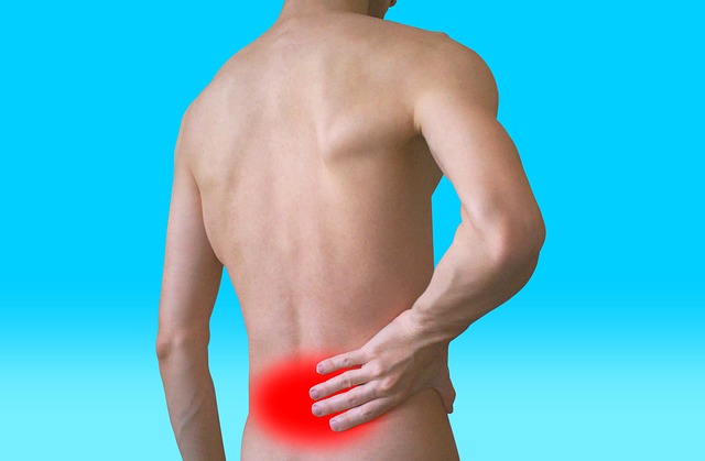
low back pain, better known as sciatica O lumbago, can have a lot of different causes, and, as a general rule, has to do with more than one. Some of the most frequent are the insufficiency of muscles in the back, overload or imbalance, as well as herniated disc, poor spinal alignment including scoliosis, a degenerative disc disease, trauma, infections or osteoporotic compression fractures, among others. This makes many people wonder if you really need a lumbar resonance.
The answer to this question is that not always necessary. This causes doubts regarding the imaging tests that should be done in case of suffering from sciatica or low back pain.. For this reason, we are going to explain what you should know about the main imaging tests most requested by spine surgeons..

Index
The imaging tests most requested by spine surgeons
X-rays
The x-rays give ideal to the spine professional. They are the best imaging test for evaluating spinal alignment and deformities.. It should be the first test used for evaluation of the spine. They are the most used when they are carried out under load, that is to say, with the patient standing, but sometimes they are performed in flexion, in extension, lying face up or face down to check the stability of the spine.
Radiology is not a painful test, but it must be taken into account that irradiation with X-rays is harmful, Therefore, it is advisable to limit its use to cases in which its use is really justified.. It should be noted that radiography of the lumbar spine involves irradiation 150 times higher than a chest x-ray.
Magnetic resonance
The magnetic resonance It is excellent for studying the soft tissues. It is able to detect disc herniation, canal stenosis, nerve or spinal cord injuries, fractures or tumors.
What can cause back pain due to alterations or inflammation in the area of the injury lumbar resonance the patient is not exposed to any type of dangerous radiation and it is also not painful. Needed, Nevertheless, Have the patient lie still for a few 15 minutes in a small space, so it can become an unpleasant situation, especially for those who have a tendency to claustrophobia.
Nevertheless, It must be taken into account that different studies have indicated that magnetic resonance imaging is not as reliable as it was considered in the diagnosis of back ailments.. Other studies show that in a lumbar resonance, until the 30% of healthy and pain-free people show herniated discs, and the 70% protrusions.
Computed tomography
The computed tomography o scanner is very useful to analyze the alignment of the spine and to study the bone, especially when the patient has had a trauma to his spine. The scanner allows to see the bone better than the MRI.
It is the best test to be able to make an evaluation of how the bone is formed after spinal surgery.. The scanner is not a painful test but it is not harmless either, since it causes the patient to be exposed to considerable irradiation, which is equivalent to several x-rays at the same time.
Lumbar MRI to complement radiography
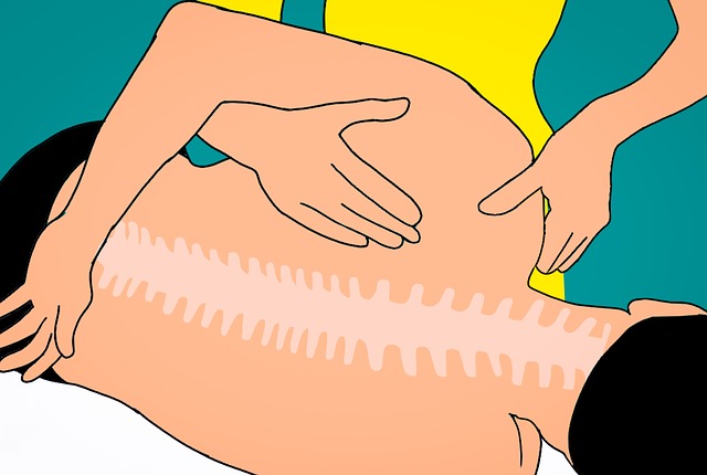
The lumbar resonance It is a non-invasive test with which to diagnose problems or diseases in the back.. It is the viable option when radiography is not enough to locate the cause of the patients' pain. It is used to obtain clear images of the lower back region.
With the lumbar resonance bone injuries can be diagnosed, as well as in intervertebral discs and ligaments, as well as diseases of the spine, like tumors, ecoliosis, tumors, infections and inflammatory diseases. Due to this, magnetic resonance imaging is the most accurate test for the diagnosis of the two most common lumbar disorders in patients of different ages., sciatica and low back pain.
With the images obtained with the lumbar resonance it is possible to identify the origin of the pain radiating along the sciatic nerve. This is used for sciatica or radiating low back pain. What's more, Through the MRI of the lumbar spine, it can be checked if there is any alteration in the spine responsible for the pain..
At the same time, when imaging the entire lower back,. This diagnostic test also makes it easier to identify other disorders that affect bowel or bladder control..
How lumbar MRI is performed
In MRI machines, a radiofrequency pulse to a tissue subjected to a magnetic field, a fabric that will be, in this case, located in the lower back. At the same time, a receiving antenna is responsible for collecting the weak signal emitted by the tissues to be examined, a signal that is transformed into an image.
To make a magnetic resonance in the back it is necessary that the person lies face up on the stretcher enabled for it, inside the MR machine. This protocol is more comfortable and less burdensome for patients in open MRI equipment, since these allow the patient to observe the outside while the test is performed, thereby reducing the level of anxiety.
Secondly, the open MRI machines high field guarantee better results. This is because the quality of the image depends, to a large degree, Make sure patients are as still as possible. This will make it easier to get a good result on these devices., especially in the case of children, that thanks to these open MRIs they can be in the company of their parents. It also helps all those patients who are clautrophobic or overweight.
The lumbar resonance, as usual, has a duration between 10 and 20 minutes, a procedure that is, Thus, pretty fast.