

Microcephaly is a developmental pathology of the skull and brain accompanied by mental retardation and neurological abnormalities.
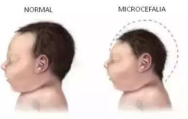

The skull is abnormally small, accompanied by low weight and underdevelopment of the brain. In this case, body proportions are absolutely normal.
It is characterized by early closure of the cranial sutures and closure of the fontanelle, convulsive syndrome, delayed motor development, intellectual defect, underdevelopment or lack of speech.
Microcephaly occurs in equal proportions between boys and girls. With a frequency of 1 case for each 10.000 kids.
Here we will explain the causes, the symptoms, the diagnosis, the most common treatments for microcephaly and their prevention.
Index
Causes of microcephaly
This pathology may have a genetic origin, caused by WDR62 gene mutation. As a result, the development of a specific protein is violated, microcephalin.
The abnormality can be triggered by a number of factors: as a result of exposure to harmful factors in the early development of the fetus and due to brain damage in the terminal stages of intrauterine development, as well as in the process of delivery and in the first months of the baby's life.
The most common causes that can be identified are:
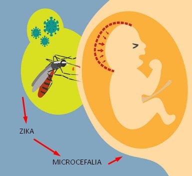
- Viruses and infectious diseases of the mother such as Zika or toxoplasmosis
- Intrauterine infections (measles, mumps, herpes, rubella, cytomegalovirus, etc.)
- Endocrine system failures in pregnant women (diabetes, hyperthyroidism)
- Exposure to antibiotics
- Genetic disorders
- Effects of radiation and bad ecology
- As a component of many inherited diseases (Payne syndrome, Giacomini syndrome)
- Metabolic disorders
- Intracranial birth injury
- Chromosomal abnormalities: Down's disease, Patau's syndrome, Edwards syndrome, síndrome “grito de gato”

In some cases, microcephaly has causes that sometimes cannot be determined, it is evident in the children of healthy parents.
Symptoms
The main symptomatic manifestation of microcephaly is a small head, disproportionate compared to the baby's body. The bevelled forehead is also observed, protruding ears and brow ridges.
As children grow up with that disease, especially from the first year of age, signs of microcephaly are hard to ignore. Some general manifestations of this disorder are:
- Small or completely closed fontanel
- Whether the fontanel was opened at the time of birth, it closes quickly, one month after delivery
- Low muscle tone
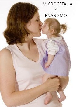
- Weight and height of the child under the norm (enanismo)
- Lack of coordination
- Seizures and loss of consciousness (Not in all cases)
- Squint
- Articulate language absent
- Absence of twists and tertiary grooves, in the structure of the brain
- General slowdown in development. Children learn late to hold their heads, turn around, sit down, crawl and walk.
- Intellectual deficiency

Diagnosis

The diagnosis of microcephaly can be made prenatally or after birth. During pregnancy ultrasound studies are performed, to compare biometric parameters in the fetus.
Ultrasound can detect small abnormalities and dimensions of the child's brain. Unfortunately this diagnosis can be made during the week 27 and 30 of pregnancy with a sensitivity of 67%.
It is because of that, if there is a suspicion of microcephaly, that is connected to a genetic or chromosomal abnormality, ultrasound detection methods should be complemented with some invasive prenatal diagnosis What: cordocentesis, amniocentesis, chorionic villus sampling and fetal karyotype.
If there is a suspicion or a family history of microcephaly, a medical history should be taken along with full parental evaluations. Where you will have genetic tests, CT scan and MRI of the head.
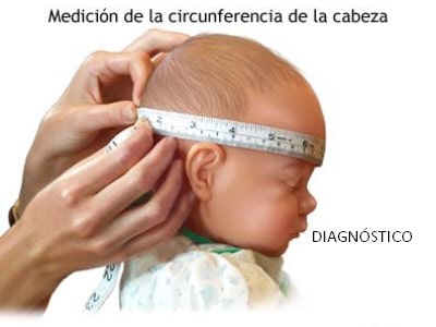
After birth, the diagnosis of microcephaly is confirmed through a visual inspection of the newborn.
To determine the extent and prognosis of the abnormality, tools such as: ecoencefalograma, electroencephalogram, magnetic resonance, CT scan and x-ray examination of the skull.
Patients with microcephaly, depending on temperament, can be divided into 2 groups: Patients in the first group are fussy, very mobile. Patients in the second group, Conversely, they are apathetic, boards, indifferent to the environment.
Microcephaly treatments
With microcephaly, the main treatment is aimed at the symptomatic support of the patients. The use of regular medication improves metabolic processes in brain tissue, by administering vitamin complexes, anticonvulsants and sedatives.
Rehabilitation in children with microcephaly includes occupational therapy, massages and physiotherapy. The treatment is aimed at the physical-intellectual development of the child and its possible social adaptation.
These methods are applied in specialized training centers to stimulate the normal course of metabolic processes in the brain..
Patients with microcephaly should be supervised by a pediatric neurologist and a pediatrician.
At the same time, the child's parents play an important role in rehabilitation. Microcephaly requires treatment and developmental therapy (memory exercises, attention, sensory stimulation, etc.
Prevention mechanisms


Prevention of microcephaly consists of careful planning of the pregnancy. Preventive exams such as the TORCH profile should be performed, CRP and prenatal fetal protection.
In the case of early intrauterine detection of microcephaly, it is necessary to decide the possibility of an artificial termination of pregnancy.
To assess the potential risk of microcephaly in subsequent pregnancies in families with a history of this condition, genetic medical counseling should be carried out.
conclusion
Microcephaly is a condition in which a child is born with a small head or the head stops growing after birth. It is a rare condition, because a child of several thousand children is born with microcephaly.
The way to determine microcephaly in a child is to measure the circumference of their head 24 hours after birth and compare the result with the WHO standard indicators for child development.
Children born with microcephaly, as they grow, may have seizures, as well as physical disabilities and learning disabilities.
There is no special treatment for microcephaly.